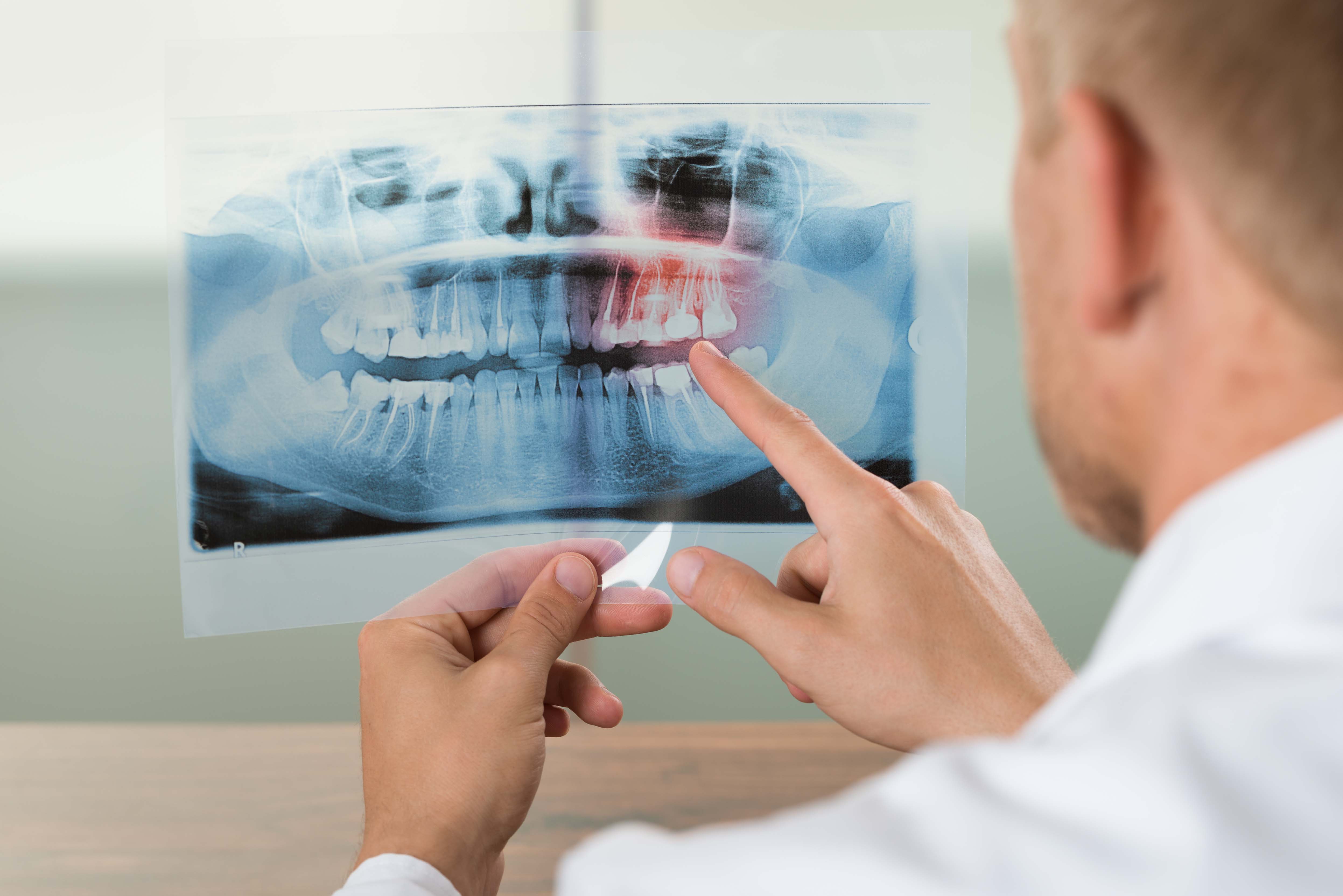
Bone cavitations can be diagnosed by x-rays, kinesology, ultrasonics and electroacupuncture. Should such a diagnosis be made treatment of the area is usually fairly straightforward. Access to the area is made by lifting back the gum and removing the hard outer cortical bone. Curettes are then used to remove all softened areas of cancellous (inner) bone.
The area is then irrigated with plain anaesthetic and sutured closed.
Healing is usually better than extractions.
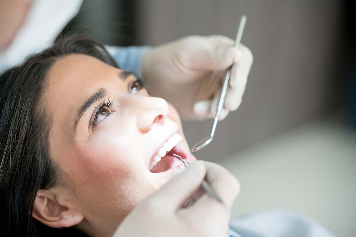
Performing a gingivectomy is quick and relatively painless. Firstly a local anesthetic is applied to completely numb the area of the gums. A small incision is made and the excess gum tissue is removed using a dental tool. A periodontal dressing covers the teeth and gums post-surgery to protect them while fully healing. This takes a few weeks, but after that the gum is completely free of pain.

A mercury amalgam tattoo is a bluish discolouration created when a piece or pieces of amalgam drop into bone or cuts in the gum during extractions or fillings and become submerged.
They are perhaps my biggest cause of failure with mercury detoxification. It is clear that all amalgam needs to be removed to enable the body to release its mercury stores. The body makes no distinction between amalgam in teeth or in the gum.
Unfortunately small amounts rarely show on x-rays and, if more than several millimeters deep, do not show in the gum. I have noticed that they do often surface 3-4 years post amalgam removal. If I see them at a check-up we remove them (and start the mercury detoxification process again).
Removal requires surgical excision. Most are very minor, yet some require extensive bone curettage.
They are essential to remove to facilitate mercury detoxification.
In 1990 in my first year of this work I was treating a lady for cavitations/osteitis. She had no teeth for 34 years and in my inexperience did not consider mercury to be an issue. Whilst lifting back the gum I noticed a blue spot and thought amalgam. I excised it and proceeded with the osteitis curettage, giving the ‘blue spot’ no more thought.
The following day the patient rang with severe symptoms of unmanaged mercury detoxification. I then remembered the tattoo and realized it was her last amalgam. I placed her on the detox program and she was improving within 24 hours and better than ever in a week. In other words one small piece of amalgam had kept all the body stores of mercury intact for 34 years.
P.S. And the dentist learned that every bit of amalgam counts!
Removing the periodontal ligament post extraction
We get many people asking us do we remove all the periodontal ligament post extraction. The answer is no. The guideline that it be completely removed is ubiquitous on the internet and has been an article of faith to many since at least 1989 when I first read it. None of the doctors who taught me have advocated total removal of the ligament and I have not done so since 1990.
What I do is remove much of it. The aim being to damage the bone in the socket (so that it remodels and repairs which then removes “toxins”). I also wish to make the area bleed so the socket fills with blood to allow socket repair and more importantly to allow the immune system to act on the area and any toxic material.
Removing the whole ligament has conse quences. In the aesthetic zone (areas you can see) it will create unsightly hollow areas above the teeth. In the lower molars you risk nerve damage that can create indefinite numbness or pain. In the upper molar area you can easily perforate the sinus.
The statement that all must be removed is a myth with big possible consequences. Like most things a middle path (partial removal) is optimal.