Not So Good Vibrations

by Shilpa Rahangdale, MD, Lisa Campana, BS, and Atul Malhotra, MD
Brigham and Women’s Hospital, Division of Sleep Medicine, Boston, MA
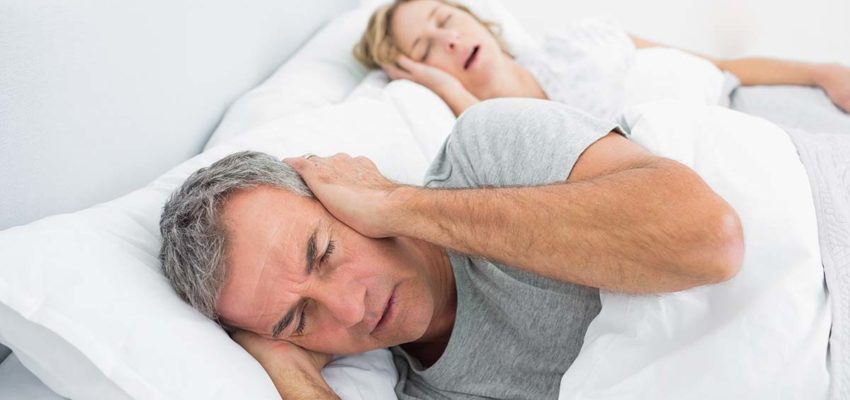
Snoring often coexists with obstructive sleep apnea (OSA) and has been used as a surrogate marker of OSA in epidemiological studies. In this issue of Sleep, Lee et al. report a link between snoring and arterial plaque in the carotid, but not femoral, arteries of patients with minimal OSA. Moreover, this association persisted after adjustment for age, sex, smoking history, and hypertension. The authors hypothesized that snoring in and of itself may not be benign, but may be responsible for focally increased atherosclerosis in the carotid arteries. If true, snoring may be elevated in importance from a social annoyance to a vascular risk factor.
Both OSA and snoring have been associated with stroke risk in various observational studies. Cross-sectional data from the Sleep Heart Health Study (SHHS) revealed an increased prevalence of carotid plaque in OSA patients, consistent with a link between OSA and stroke. Prospective follow-up of OSA patients in SHHS demonstrate that OSA was independently associated with stroke (at least in some subgroups), further supporting the association between OSA and stroke (as yet unpublished). Additionally, OSA may preferentially favor the development of carotid atherosclerosis, based on a stronger association with stroke than coronary disease from SHHS. One major implication of these findings is that OSA effects may have been more robust in these prior studies if the affected patients were compared with controls without snoring (i.e., morbidity from snoring in the control groups may bias towards the null hypothesis). Regardless, the link between snoring and OSA (which is usually accompanied by snoring) with stroke appears strong, and OSA patients may be more likely to develop carotid disease.
Primary snoring, previously thought to be benign, is associated with potentially deleterious pathophysiological changes beyond tissue vibrations. Snoring results from tissue vibrations in the upper airway due to turbulent flow, implying flow limitation. Inspiratory flow limitation is accompanied by markedly negative intrathoracic pressure swings, which increase left ventricular transmural pressure with associated increases in left ventricular wall tension and afterload. Additionally, snoring (depending on the hypopnea definition used) may be associated with increased arousals and catecholamine surges. Primary snoring, not just OSA, is associated with daytime sleepiness. Flow limitation may lead to hypercapnia, which can stimulate catecholamine release causing vasoconstriction. Thus, snoring, even in the absence of apneas and hypopneas, can cause cardiovascular consequences and daytime sleepiness in addition to inspiratory vibrations.
Snoring may be more likely to contribute to carotid atherosclerotic damage in part due to these inspiratory vibrations. The vibrations produced by snoring are hypothesized to initiate plaque formation (and possibly embolization from existing plaques) by the transmission of pressure waves to surrounding tissues, promoting vascular damage. In rabbit models of snoring, vibration produced in the upper airway lumen transmitted energy (pressure waves) to the peripharyngeal tissues, the carotid artery wall, and the carotid artery lumen. In addition, rat models of snoring are associated with inflammation of upper airway tissues, including upregulation of NF-κB and MIP-2 in the tissues. This concept is also supported by in vitro data suggesting increases in inflammatory markers in bronchial epithelial cells exposed to 60-Hz vibration. Additionally, abnormal sensory nerve function in the upper airway in OSA patients has been observed. Nguyen and Kimoff have noted that both nonapneic snorers and OSA patients had impaired upper airway sensation compared to non-snorers. Thus, snoring related vibrations increase upper airway inflammation and may affect pharyngeal sensory nerve function.
Although there is minimal literature on the effect of snoring-induced vibration on the vasculature or in endothelial cells in vitro, there exists clinical and experimental evidence suggesting that vibration can damage the vasculature. Workers exposed to vibrating machines may develop Raynaud’s syndrome, characterized by episodic digital ischemia with microvascular spasm triggered by cold or emotional stress. Kennedy et al. demonstrated elevated inflammatory markers (serum ICAM-1 level) and impaired smooth muscle response in the skin blood vessels at the fingertips of people who were occupationally exposed to chronic vibration. In animal models, vibration of the rat tail artery damages both endothelial cells and smooth muscle cells of the vasculature by causing a combination of intense vasoconstriction and mechanical stresses. Vibration appears to disrupt the endothelial tight cell junctions, which normally isolate the interstitium osmotically from the blood. This change may allow leakage of fluid into the subendothelial space, promoting increased hydrostatic pressure and disruption of other cell-cell and cell-matrix interactions. Thus, vibration can cause injury and inflammation in both vascular endothelium and smooth muscle.
In summary, Lee et al. present a novel concept with plausible biological mechanisms and potential clinical implications. If snoring-induced vascular damage is a real entity, one wonders if therapy for primary snoring is warranted to treat vascular risk. In addition, although snoring is generally eliminated by continuous positive airway pressure (CPAP), one may argue that particular care is needed to titrate CPAP levels to eliminate snoring. Finally, given the very high population prevalence of snoring, prioritization of patient groups at risk of snoring-induced complications who should be targeted therapeutically may be important.
Many important investigative questions also remain. How should snoring be assessed? By time (as done by Lee et al.)? By vibration frequency? And/or by amplitude? Other important research questions include: How does vibration affect endothelial cell function? Is this effect different in the large vessels as opposed to the microcirculation? Can these snoring induced vascular complications be blocked pharmacologically (e.g., β- or calcium channel blockers and/or statins)? Do the femoral and carotid arteries have other biological differences apart from proximity to the airway that may explain the new findings? At this time, the concept of snoring induced atherosclerosis, while thought provoking, still requires further study prior to changes in clinical practice. We applaud Lee et al. on their intriguing findings.

REFERENCES1. Malhotra A, White DP. Obstructive sleep apnoea. Lancet. 2002;360:237–45.2. Lee S, Amis T, Byth K, et al. Heavy snoring as a cause of carotid artery atherosclerosis. SLEEP. 2008;31:1207–13.3. Yaggi H, Mohsenin V. Obstructive sleep apnoea and stroke. Lancet Neurol. 2004;3:333–42.4. Yaggi HK, Concato J, Kernan WN, Lichtman JH, Brass LM, Mohsenin V. Obstructive sleep apnea as a risk factor for stroke and death. N Engl J Med. 2005;353:2034–41.5. Arzt M, Young T, Finn L, Skatrud JB, Bradley TD. Association of sleep-disordered breathing and the occurrence of stroke. Am J Respir Crit Care Med. 2005;172:1447–51.6. Wattanakit K, Boland L, Punjabi NM, Shahar E. Relation of sleep-disordered breathing to carotid plaque and intima-media thickness. Atherosclerosis. 2008;197:125–31.7. Shahar E, Whitney CW, Redline S, et al. Sleep-disordered breathing and cardiovascular disease. cross-sectional results of the Sleep Heart Health Study. Am J Respir Crit Care Med. 2001;163:19–25.8. Hosselet JJ, Norman RG, Ayappa I, Rapoport DM. Detection of flow limitation with a nasal cannula/pressure transducer. Am J Respir Crit Care Med. 1998;157:1461–7.9. Malhotra A, Muse VV, Mark EJ. Case records of the Massachusetts General Hospital. Weekly clinicopathological exercises. Case 12-2003. An 82-year-old man with dyspnea and pulmonary abnormalities. New Engl J Med. 2003;348:1574–85.10. Fessler H, Brower R, Wise R, Permutt S. Mechanism of reduced LV afterload by systolic and diastolic positive pleural pressure. J Appl Physiol. 1988;65:1244–1250.11. Gottlieb DJ, Yao Q, Redline S, Ali T, Mahowald MW. Does snoring predict sleepiness independently of apnea and hypopnea frequency? Am J Respir Crit Care Med. 2000;162:1512–17.12. Edwards N, Wilcox I, Polo OJ, Sullivan CE. Hypercapnic blood pressure response is greater during the luteal phase of the menstrual cycle. J Appl Physiol. 1996;81:2142–6.13. Amatoury J, Howitt L, Wheatley JR, Avolio AP, Amis TC. Snoring-related energy transmission to the carotid artery in rabbits. J Appl Physiol. 2006;100:1547–53.14. Almendros I, Acerbi I, Puig F, Montserrat JM, Navajas D, Farre R. Upper-airway inflammation triggered by vibration in a rat model of snoring. Sleep. 2007;30:225–7.15. Nguyen AT, Jobin V, Payne R, Beauregard J, Naor N, Kimoff RJ. Laryngeal and velopharyngeal sensory impairment in obstructive sleep apnea. Sleep. 2005;28:585–93.16. Kennedy G, Khan F, McLaren M, Belch JJ. Endothelial activation and response in patients with hand arm vibration syndrome. Eur J Clin Invest. 1999;29:577–81.17. Curry BD, Govindaraju SR, Bain JL, et al. Evidence for frequency-dependent arterial damage in vibrated rat tails. Anat Rec A Discov Mol Cell Evol Biol. 2005;284:511–21.
This article is provided here courtesy ofAssociated Professional Sleep Societies, LLC
David Howard has been practising Holistic Dentistry since 1990 when he ceased doing Root Canals and Amalgam fillings. Many years of study and experience have made him a leader in the field and a safe pair of hands for the patient intending to shift from the old to the new paradigm of dentistry.
Latest posts by Dr David Howard (see all)
- Root Cause by Netflix - February 17, 2019
- Mercury – I Should be Deranged or Dead - January 23, 2019
- Snoring - January 23, 2019