Toxic Teeth: The Chronic Mercury Poisoning of Modern Man

Introduction
Exposure to mercury from ‘silver’ dental fillings has gained considerable notoriety in the general media during the past decade. Specific attention has focused on the potential consequences for human health and the general well-being of the global environment, The modern ‘silver’ filling has been used as the main tooth restorative material for over 180 years and presently accounts for 75-80% of all tooth restorations. These ‘silver’ fillings contain about 50% mercury by weight, 35% silver, 13% tin, 2% copper and a trace of zinc. (2)
Each tooth restoration has a mercury mass of 750-1000mg and should more properly be called a mercury filling. They have a functional life of about 7-9 years, after which they are usually replaced with another mercury filling. (3.4) Hundreds of metric material, as particulate waste from the dental office, finds it’s way into the sewerage and refuse systems.
Within the dental profession, the issue of mercury filling safety has recurred cyclically, After the introduction of the modern dental amalgam in 1812 by Joseph Bell, a British chemist, a ‘silver paste’, which was a combination of silver fillings from coins and mercury, became fashionable for tooth restoration, Since the coins were not pure, expansion of the material resulted in tooth fracture and/or ‘high bite’. In America during the 1800’s, concern about the possible mercury toxicity caused the American Society of Dental Surgeons to make mercury usage an issue of malpractice, mandating that its members sign an oath not to use mercury-containing materials; However, use of mercury fillings increased because it offered dentists an economic advantage. The fillings were also user friendly and durable in the mouth. By 1856, the American Society of Dental Surgeons we forced to disband because of dwindling membership over the mercury filling issue. In its place rose the American Dental Association, founded by those who advocated silver amalgam – mercury use in dentistry. (5-7) Again in the 1920s, a controversy erupted after a publication of articles and letters by German chemistry professor, Alfred Stock, who attacked mercury filling usage for possible toxic effects. (8-13) That debate abated and the dental profession’ opinion still remains unchanged.
Today, 182 years later, the American Dental Association has amended its code of ethics to make the removal of serviceable mercury fillings an issue of unethical conduct, if the reason of removal is to eliminate a toxic material from the human body and if this recommendation is made solely by the dentist. (14) In the Association’s view, a dentist is ‘ethical’ to place the mercury material and recommend its safety. However, of the dentist suggests that mercury can result, them the dentist is acting, ‘unethically’. Clinically serviceable mercury fillings can be ‘ethically’ removed if done for aesthetic reasons, at the request of a physician, or at the patients request (without prompting).

Mercury release from dental fillings
Mercury vaporises continuously from dental fillings, and this is intensified by chewing. (15,16) tooth brushing (170 and hot liquids). (18) After mastication or even decline to the lower pre-chewing level. (16) Also the greater the number of fillings and the larger the chewing surface area, the larger the mercury exposure. (15-16) Thus, the average individual us on a roller coaster of mercury vapour exposure during the day. Breakfast will cause the release rate to increase and just as the rate us slowing again it is time for the mid-morning coffee break. Lunch, mid bedtime all contribute to the daily exposure to mercury from dental fillings.
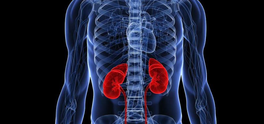
It is estimated that the average individual, with eight biting surface mercury fillings, is exposed to a daily dose uptake of about 10ug mercury from their fillings. (19) Select individuals may have daily doses 10 times higher (100ug) because of factors which exacerbate the mercury vaporisation. These factors include frequency of eating, chronic gum chewing, chronic tooth grinding behaviour (usually during sleep), the individual’s chewing pattern, consumption of hot foods and drinks, and mouth and food acidity, (16) Corroborating human autopsy evidence (20-22) showed that the brain and kidney tissues contained significantly higher amounts or mercury in individuals who had mercury fillings. Furthermore, the concentration of mercury in the brain in the subjects with mercury fillings correlated with the number of fillings present.
Historically, the espoused opinion of dentistry insists that, once mixed, the mercury is locked into the fillings, (23) The above body of experimental evidence suggests that this opinion is totally without merit., Despite the replicated research findings, many national dental trade associations still claim that mercury fillings are safe. (24)
They base their conviction on the anecdotal facts that mercury fillings have been used for over 150 years, billions of fillings have been placed, and they do not see sickness or death from mercury exposure. (25) But, the diagnosis of mercury toxicity lies outside the scope of dentistry, falling more appropriately within the jurisdiction of medicine. Dental institutions do not have scientific expertise or the resources to undertake the necessary studies to resolve this issue scientifically. Thus, mercury fulling safety has not been suitably addressed until recently, when academic medicine became aware of this insidious exposure to the element. From the medical perspective, dental amalgam fillings are a significant mercury source, having potential medical consequences.

Tissue uptake of mercury from dental fillings
Recent investigations in sheep and monkey animal models demonstrate that dental mercury accumulates in all tissues of the adult, and us at its highest in the kidney and liver. This accumulation is so extensive that it can be visualised on a whole-body image scan. (26-27) Research also shows that a high level of dental amalgam mercury in monkey kidney is still present one year after the filling us placed (28). These prospective studies confirm the retrospective human autopsy studies discussed previously (20-22). Also, mercury from dental amalgam will cross the placenta and begin accumulating in the developing foetus within two days of the filling’s installation in pregnant sheep (29).
Here it is highest in the foetal liver rather then the kidney. The mother’s milk also showed evidence of mercury, suggesting that the newborn would have an additional exposure to the element (29).
Recent human chelation studies show an association between urinary mercury excretion and the presence of mercury fillings. (30-33) For example, one study showed that, after chelation challenge with DMPS (2,3-dimercaptopropane-1-sulphonate), urinary mercury excretion is significantly higher from subjects with mercury fillings that from those without. It was concluded that at least two-thirds of the excreted mercury, after the DMPS challenge, originated from dental restorations.(30)
On the basis of this research, there is now an international scientific consensus that the mercury from dental tooth restorations constitutes the largest non-occupational source of mercury in the general population, being greater that all other environmental sources combined! (34-36) Yet, the dental profession still insists, without evidence, that the exposure is insignificant and has no potential to produce harm.

Pathophysiological consequences
During the past few years, medical research has demonstrated a relationship between mercury exposure and pathophysiology in various animal models. In sheep exposed to mercury from in “in situ” tooth fillings, kidney function was impaired. After 30 days of chewing, the sheep lost half of their kidney filtration ability, they began to have difficulty regulating sodium and they demonstrated a reduced albumin excretion. Control sheep treated with non-mercury dental fillings did not show such effects. (37) A study of ten humans with mercury fillings, showed that the plasma mercury level dropped by half and the urinary mercury level declined by a quarter over the year after the fillings were removed compared with the pre-removal level. Most notable was the finding that a year after the fillings were removed, the urinary albumin level was significantly higher than that four months before removal. (38) Albumin, a common protein found in the blood, has a molecular size that means in a normal, healthy kidney a specific amount will get filtered and passed into the urine. In the sheep, the placement of mercury fillings caused a fall in the urinary albumin, signifying kidney pathophysiology- either a reduced ability of the membrane (filter) to function properly or a fall in blood pressure in the filtering area. In humans, the removal of mercury fillings results in an elevation in urinary albumin, indicating a kidney homeostatic readjustment towards normal function. The agreement between the sheep and human data is remarkable.
A recent collaborative paper between three North American universities used monkeys to show that oral and intestinal bacteria (for example, streptococci, enterococci and enterobacterieae) exhibit a significant increase in mercury and anitibiotic resistance within two weeks of cannot be mercury filling placement. (39) The mercury-resistant bacterial species had resistance to various antibiotics such as ampicillin, tetracyclines, streptomycin, kanamycin, erythromycin and chloramphenicol.
They had not demonstrated such resistance before placement. This is the first direct experimental confirmation of a non-antibiotic resistance, It occurs because in some bacteria mercury-resistance and antibiotic-resistance are encoded in adjacent small genetic sites within plasmids. (40) When exposed to environmental mercury, this genetic material is activated to protect the bacteria from the lethal mercury. The plasmid is also replicated and passed on to other bacteria, ensuring species survival. In so doing, the antibiotic resistance also spreads to the other bacteria.
Antibiotic resistance is an important issue in medicine today.(41) It has been estimated that 80% of mercury-resistant bacterial strains also show an increased resistance to one or more conventional antibiotics. Thirty percent of all hospitalised patients in North America receive antibiotic therapy. (42) and antibiotics made up a tenth of the total $5.1 billion drug sales in Canada during 1992. (43) Moreover, ten of the top 20 generic drugs prescribed during 1990 in the US were antibiotics. (44) Yet, antibiotics appear to be losing their clinical potency and stronger antibiotic medications at increasing dosages are necessary to combat many common infections (41).
Recently, investigations have suggested that mercury may be involved in common brain diseases and that the source of the mercury is likely to be dental fillings. (45-47_ In a human autopsy study, (47) brain tissue from people with Alzheimer’s Disease t death were compared with an age-matched group of control brains from subjects without Alzheimer’s Disease. The only significant difference in metal content between the two groups of brains was mercury, being considerably higher in the Alzheimer group. The mercury concentration was prominent in the hippocampus, the amygdala and particularly in the nucleus basils, all brain structures involved in memory function. Other metals examined were not significantly different in the two groups of subjects. Dental Histories were unavailable, but other authors speculated that the likely source was mercury fillings.
The effect of mercury on the central nervous system neurone membrane integrity has also been examined. Here the mercury specifically affects tubulin, a brain neuronal dimer protein responsible for the proper microtubule formation of brain neurones. (48) Both in vivo and in vitro experiments demonstrated that mercury chelated to amino neurofibrilar tangles, which are a recognised characteristic of Alzheimer’s Disease. Inorganic mercury affects adenosine diphosphate ADP ribosylation is the rate limiting process involved in the polymerisation of tubulin and actin monomers into the structure of the neurone membrane. Most recently our laboratory has demonstrated that ionic mercury and elemental mercury vapour markedly diminish the binding of tubulin to guanosine triphosphate and thus inhibit tubulin polymerisation, which is essential for the formation of microtubule in the central nervous system. (50) These studies are direct quantitative evidence for a connection between mercury exposure and neuro-degeneration.
Other investigations have examined the mercury hypersensitivity from dental amalgam in patients with and without oral lichen planus lesions, an autoimmune disease which has oral white patches as medical sign. (51-53)
These studies showed that patient groups having oral lichen planus had a much higher incidence of mercury patch test (skin allergy testing) reactivity (16-62%) than the control groups did (38%). Removal of the mercury fillings resulted in amelioration of the oral symptoms.

Governmental regulatory action
In 1987 an ‘expert panel’ commissioned by the Swedish government concluded that mercury fillings were ‘unsuitable from a toxicological point of view’. Based on this advise, the Swedish social welfare/health department (Socialstyrelsen) announced that steps would be taken to eliminate dental amalgam usage and recommended that comprehensive mercury fulling treatment on pregnant women should be stopped to prevent mercury damage to the foetus. (54) Shortly after, the German ministry of health (BDA) issued similar advise. (55) In October 1989, the Swedish Director of Chemical Inspection (KEMI), responsible for environmental protection, declared that amalgam would be banned (56). In January 1992, the German BDA informed manufacturers of its intention to ban amalgam production (57). The BDA removed low copper non-gamma-2-amalgam from the market and published a pamphlet recommending avoiding mercury filling use in individuals with kidney disease, children to age 6, and pregnant woman (58).
In August 1992, the Swedish government suggested a timetable to phase out mercury fillings. Environmental concerns were used as the official reason for amalgam discontinuation, but the government did acknowledge the toxicological risk to patients and stated that mercury fillings should no longer be used in children by July 1993, in adolescents to age 19 by July 1995, and in all Swedish citizens by 1997 (59). The Austrian Minister for health announced that the use of mercury fillings in children would be banned in 1996 and discontinued in all Austrians by the year 2000 (60). In 1994, the Swedish Dental Association acknowledged that its leadership had previously been incorrect in its position on mercury filling safety. It now supports a discontinuation of mercury use in dentistry. Other industrialised countries, for what ever reason, appear to be side-stepping the issue.

Conclusions
As might be expected, the dental profession has not responded well to these data. Some national dental associations have attempted to influence public and governmental opinion by endorsing quasi academic symposia pervaded with amalgam advocates. These gatherings are non-consensus meetings often under government auspices where the moderators responsible for drawing the conclusions are typically inclined towards the prevailing dental orthodoxy and the conclusions reached often blatantly disregard the experimental data presented. Most damning to the dental profession is that it has not advanced any reputable experimental evidence of its own to support its belief in mercury filling safety.
The medical research evidence has been clear for some time. Dental amalgam mercury fillings – constitutes a significant source of chronic exposure to mercury in the general population. This exposure is unnecessary and cannot be justified by risk/benefit analysis. While incriminating medical research continues to be published, the dental profession persists in placing itself in the untenable predicament of advocating an anecdotal position on mercury filling safety. The mercury filling advocates can be criticised for their shortage of supporting research evidence; however, so can many mercury filling opponents, who irresponsibly go far beyond the limits of the experimental data by suggesting that miraculous cures will occur after removal of the fillings. Still, the mercury exposure from dental silver amalgam is toxicologically significant and research into its possible effects is at an early stage. Perhaps in 1,000 years from now, historians will look back and draw comparisons between the chronic lead poisoning of the Roman Empire and the insidious mercury poisoning from our toxic teeth.
Professor Vimy
Faculty of Medicine
University of Calgary, Calgary, Alberta,Canada

References
1 Baurer, J.G & First, H.A, CalifDent.Assoc, J,.1982, 10, 47-61
2 Skinner, E.W.,B& Phillips, RW,. ‘The Science of dental materials’, 6th Edition, Philadelphia: W.B.Saunders Co, 1969, 303 and 332.
3 Paterso4 N., Br Dent J 1984, 157, 23-25
4 Phillips, RW, etal, J., 1984, 157, 23-25
5 American Academy of Dental Science, ‘ A history of dental and oral science in America,’ Philadelphia: Samuel White, 1876
6 Bremmer, D.K, ‘ The story of dentistry’, Revised 3rd Edition, Broo/dyn:
7 Ring, M, ‘ Dentistry, an illistrated history’ New York: Harryu Nabrams, 1985
8 Stoek, A., Z Angew, Chemie, 1926, 39, 984-9
9 Stock, A., ibid, 1928, 41, 663-72
10 Stock,A,.Z, Inorg, Jillgem. Chemie, 1934, 217, 241-53
11 Stock, A., Naturwisch, 1935, 28, 453-6
12 Stoek, A., Arch, GewerbepathGewerbchygie, 1936, 7, 388-413
13 Stoek, A., Ber Dtsch Chem. Ges., 1939, 72, 1844 57
14 American Dental Association, ‘ Principle of ethics and code of professional conduct’, Section 1-J: Representation of care fees , 211 E Chicago Avenue, Chicago IL US, 64611
15 Vimy, M.J., & Lorscheider, F.L.,J.Dent. Res, 1985, 64, 1069-71
16 Vimy, M.J.,& Lorsheider, F.l. ibid, 1985, 64. 1072-5
17 Patterson, J.E., Weissberg, B.G., and Dennison, P.J., Bull. Environ, Contam, Toxicol, 1985, 34, 459-68
18 Fedin, B., Swed. Dent. J., 1988, 3, 8-15 Vimy, M.J., & Lorscheider, F.L., J Trace Elem. Exper. Med., 1990, 3, 111-23 aussrgen von medizin und zahnmedizin’, Syrnposium, Koln, West Germany, March 1984, Abst D29
21 Nylander, M., Friberg, L.,& Lind, B,. Swed. Dent. J,. 1987, 11, 179-87
22 Eggelston, D.W., & Nylander, M.,J Prosth. Dent., 1987, 58, 704-7
23 ADA News, Editorial and accompanying patient handout on the safety of dental amalgam, American Dental Association, 2nd January 1984
24 Truono, E.1., “Letter of importance”, J.Amer: Dent. Assoc. 1991, 122, 8-14
25 Ameican Dental Association News Release, 1990
25 Hahn. LJ, etal FASEB J., 1989, 3, 2641-6
27 Hahn. LJ, etal, ibid, 1990, 4, 3256-60
28 Danscher, G., Horsted-Bindslev, P., & Rungby, l Exp. Mol. Path,. 1990, 52, 291-9
29 Vimy, M.I., Tahhashi, Y., & Lorscheider, F.L., Amer: J.Physiol., 1990, 258, R939-R945
30 Aposhian, H.V. etal, FASEBJ., 1992, 6, 2472-6
31 Gerhard, I., Waldbrenner, P.,Thuro, H., & Runnebaum, B., Clln. Lah, 1992, 38, 404-11
32 Zander, D., Ewers, U., Freier, I., & Brockhaus, A., ZbL Hyg, Umwelt., 1992, 192, 447-S4
33 Zander, D., Ewers, U., Freier, I., & Brockhaus A, ibid, 1992, 193, 318-28
34 Clarkson, T.W., Hursh, J.B., Sager, P.R, & Syversen, T,L.M., in ‘Biological monitoring of toxic metals’ (Eds T W Clarkson, L Friberg, G F Nordberg and P R Sager) New York Plenum Press, 1988, 199-246
35 Vimy MJ., & Lorscheider, F L ,. TraceElem. Esper Med., 1990,3 111-23
36 World Health Organisation ‘Environmental health criteris 118, inorganic mercury’ Geneva: WHO 1991, 36
37 Boyd ND etal,AM, J.Physiol., 1991. 2CI. R101& R1014
38 Molin M., dal. ActaOdontol.Scand., 1990, 48, 189-202
39 Summers, A,O., etal Antimicrob. Agents and Chemother, 1993, 37, 825-34
40 Gilbert, M.P., & Sununers, A.O., Plasmid, 1988, 20, 127-36
41 Cohe4ML., Science 1992, 1050-5
42 Gilrnan, H,G. Ratt TW Nies. A S,. & Taylor. P “Goodman and Gitmans
43 Intercontinental Medical Statistics, IMS, Canada, 1992
44 Pharmacy rmes, Aprill 1991, 58
45 Khatoon, S., Campbell S.R, Haley, B.E., & Slevin, J.T., Ann. Neurol., 1989, 26, 210-5
46 Thompso4 C.M., et al, Neurotoxicology, 1988,9,1-7
47 Wenstmp, D., Ehmann, W.D., & Markesbery, W.R, Brain Res., 1990, 533, 125-31
48 Duhr, E., etal, FASEB J., 1991, 5, A456
49 Patkiewicz, P., Zwiers, H., & Lorscheider, F.L.,J. Neurochem., 1994, 62, 2049-52
50 Lorscheider, F.L., Vlmy., Pendergrass, 1.C., & Haley, B.E@ Abstract presented at the 12tb Internationat neurotoxicotogy Conference, University of Arkansas medicat Center, Hot Spring, 30tb october-2nd November, 1994
51 Finne, K, Goransson, K, @ Wmckta, L., Int. J. Oral Surg., 1982, 11, 2369
52 Lundstrom, I.M.C., ibid, 1983, 12, 19
53 Mobacken, H., Hersle, K, Sloberg, K, & Thilander, H., Contact Dennatitis, 1984, 10, 115
54 Socialstyrelsend (Sweden, Social Welfare and Health Administration). ‘Redovisar, kvicksilver/amalgam halsonsker’, Stockholm: Allanna Forlaget AB, 1987, 1032-39
55 Bundesgesundheitsamt (German Ministry of Health), ‘Machine desig 25th August 1988, 274
56 KEMI (Swedish Chemical Inspection Agency), ‘Amalgam will be banned’, Dagens Nyheter, 6th October, 1989
57 Bundesgesundheitsamt (German Ministry of Health), Letter to pharmaceutical companies, 29th January, Jlrtezein ng (Physician’s Daily), 3rd march, 1992
58 Bundesgesundheitsamt (German Ministry of Health), ‘Arnalgame nevbenwirkungen und bewertung der toxizitat’, Zahnartzt Woche (DZW), 1992, 8,1
59 Socialstyrelsen (Swedish Social Welfare and Health Administration), Press Release, 28th August 1992
60 Austrian Minister of Health, ‘Austria to be amalgam free by year 2000’, FD1 Dental World, March/April, 1993,6
61 Swedish Dental Association, Swedish News Bureau, rr, 17th January, 1994
62 Lorscheider, F.L., & Vlmy, M.J., FASEB J., 1993, 7, 1432


